 Open full image ↗
01 · Heart chamber
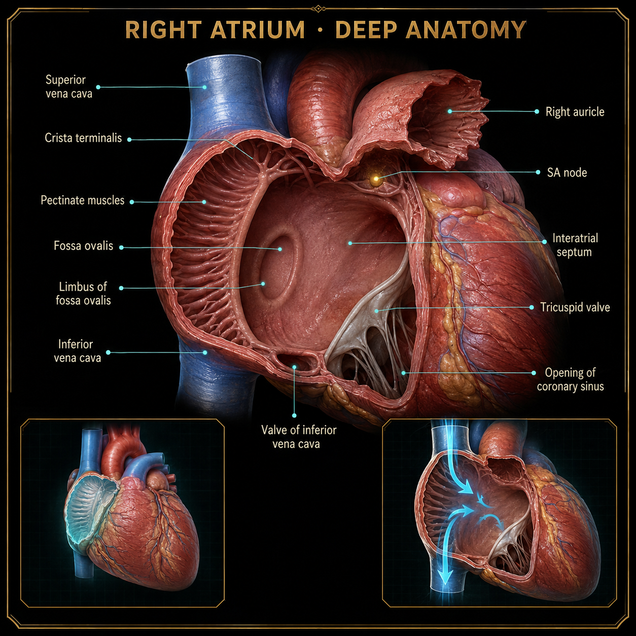
Right atrium
The receiving chamber for deoxygenated blood returning from the body through the superior and inferior venae cavae.
Body in Rhythm · visual library
Study each cardiovascular structure without the music or challenge timer. Select any plate to open the full-resolution anatomy.
Open full image ↗The receiving chamber for deoxygenated blood returning from the body through the superior and inferior venae cavae.
 Open full image ↗
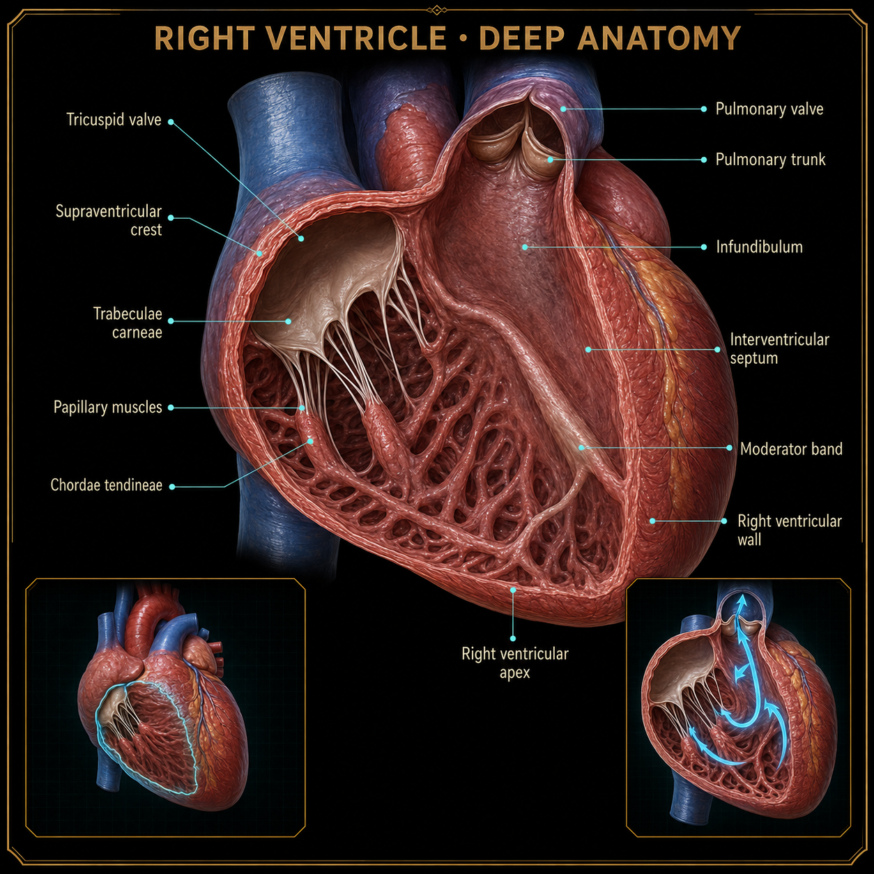
Pumps deoxygenated blood through the pulmonary valve and toward the lungs.
 Open full image ↗
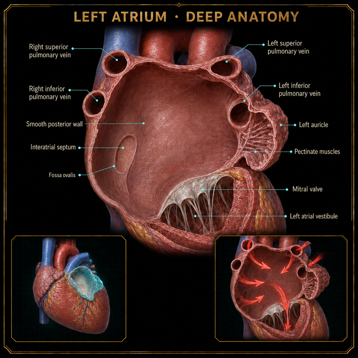
Receives oxygenated blood returning from the lungs through the pulmonary veins.
 Open full image ↗
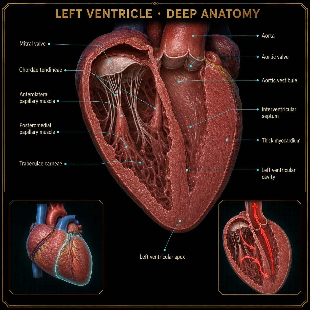
Its thick muscular wall creates enough pressure to send oxygenated blood through the entire body.
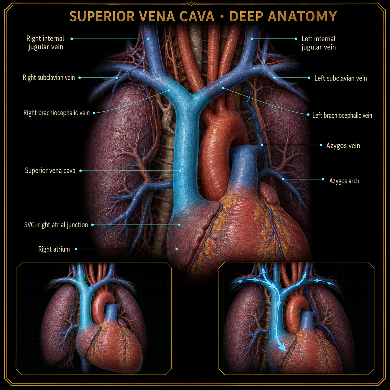 Open full image ↗
This major vein carries deoxygenated blood from the upper body into the right atrium.
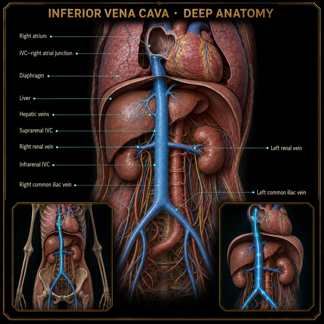 Open full image ↗
This major vein carries deoxygenated blood from the lower body upward into the right atrium.
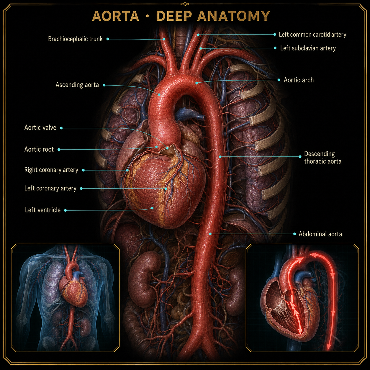 Open full image ↗
The largest artery carries oxygenated blood from the left ventricle into systemic circulation.
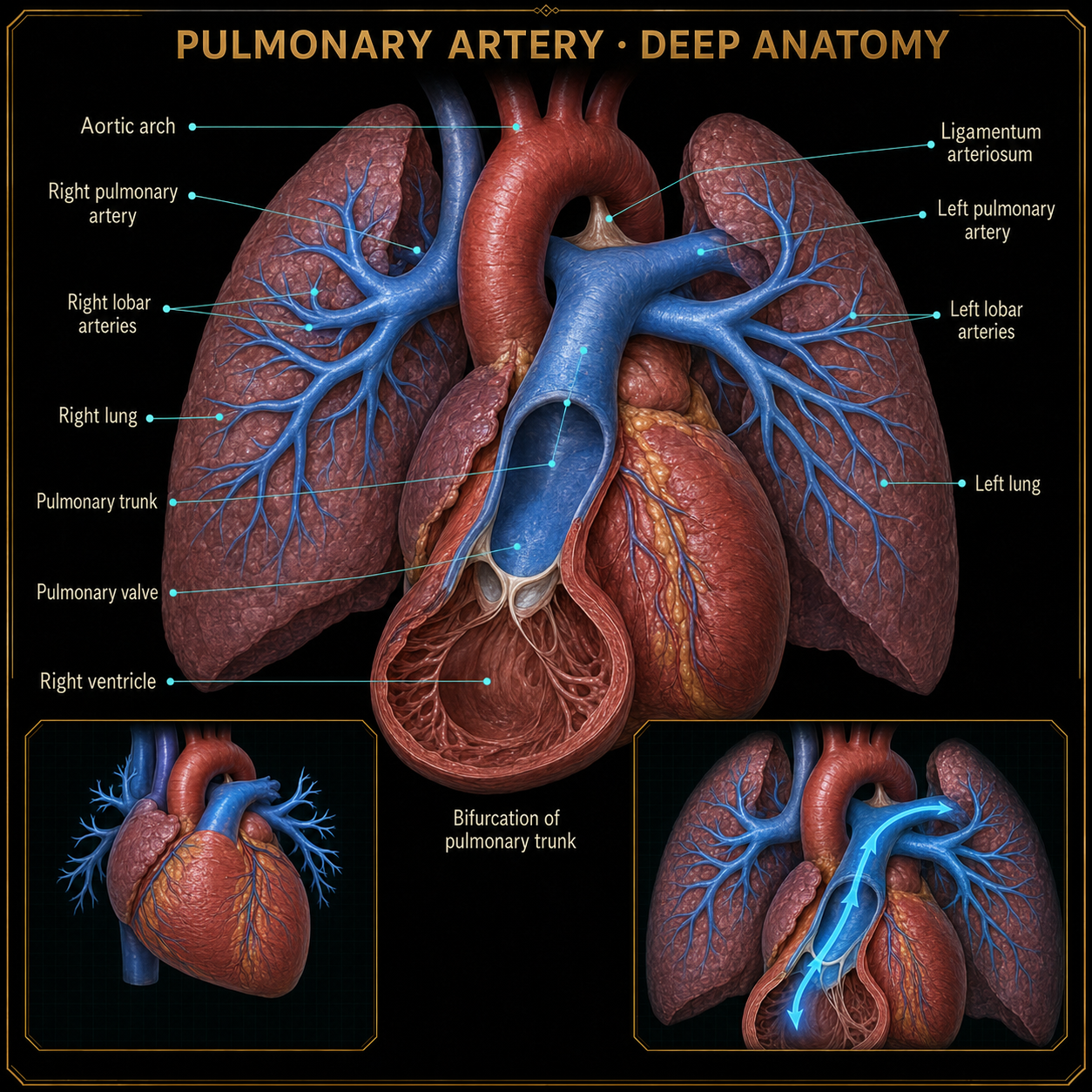 Open full image ↗
Carries deoxygenated blood from the right ventricle toward the lungs for gas exchange.
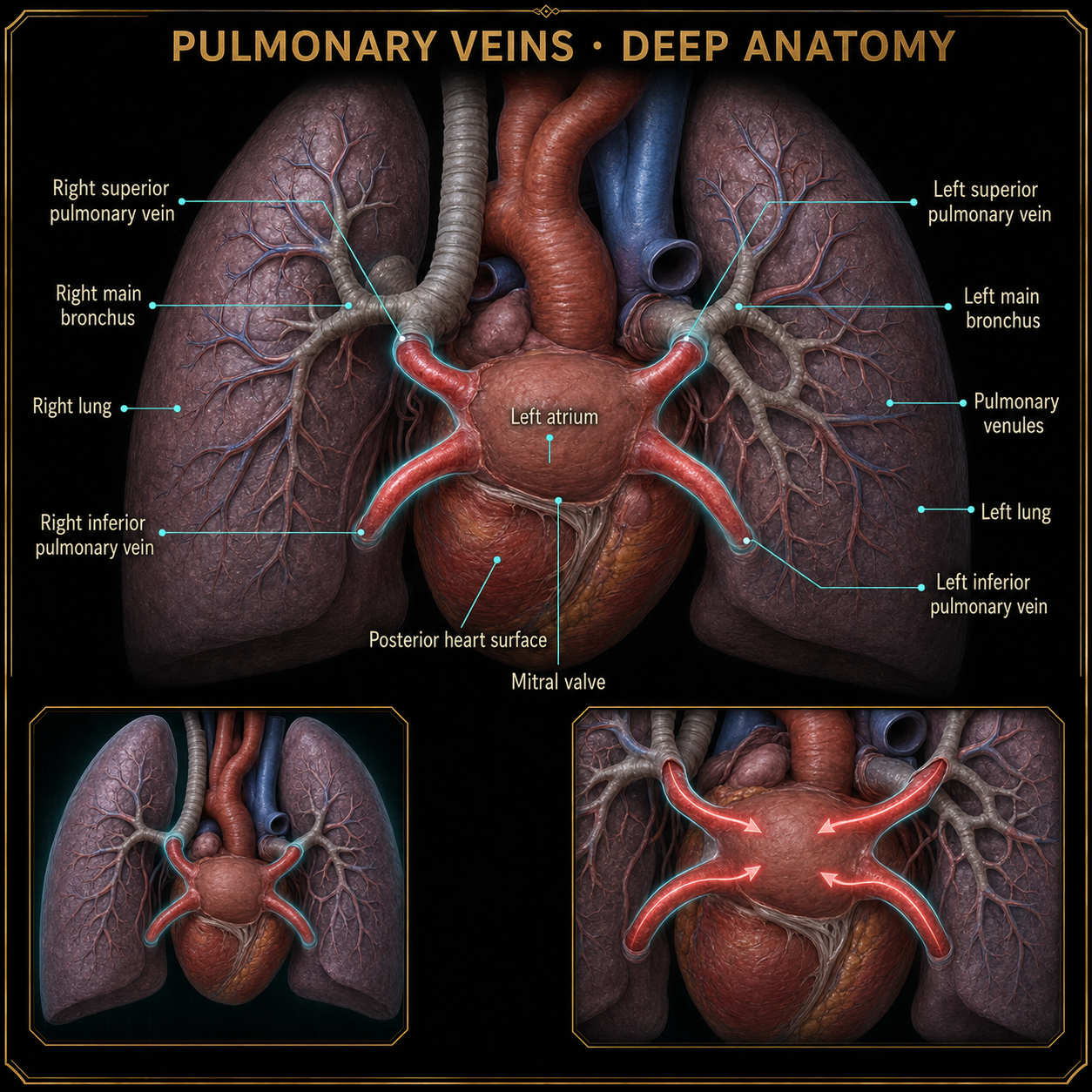 Open full image ↗
These vessels are the vein exception: they carry oxygenated blood from the lungs into the left atrium.
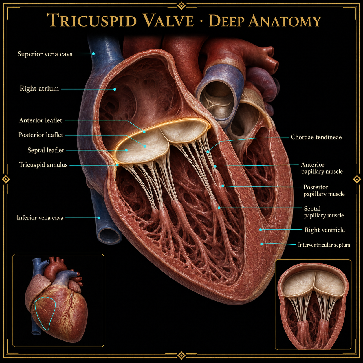 Open full image ↗
The right atrioventricular valve directs blood from the right atrium into the right ventricle and helps prevent backflow during ventricular contraction.
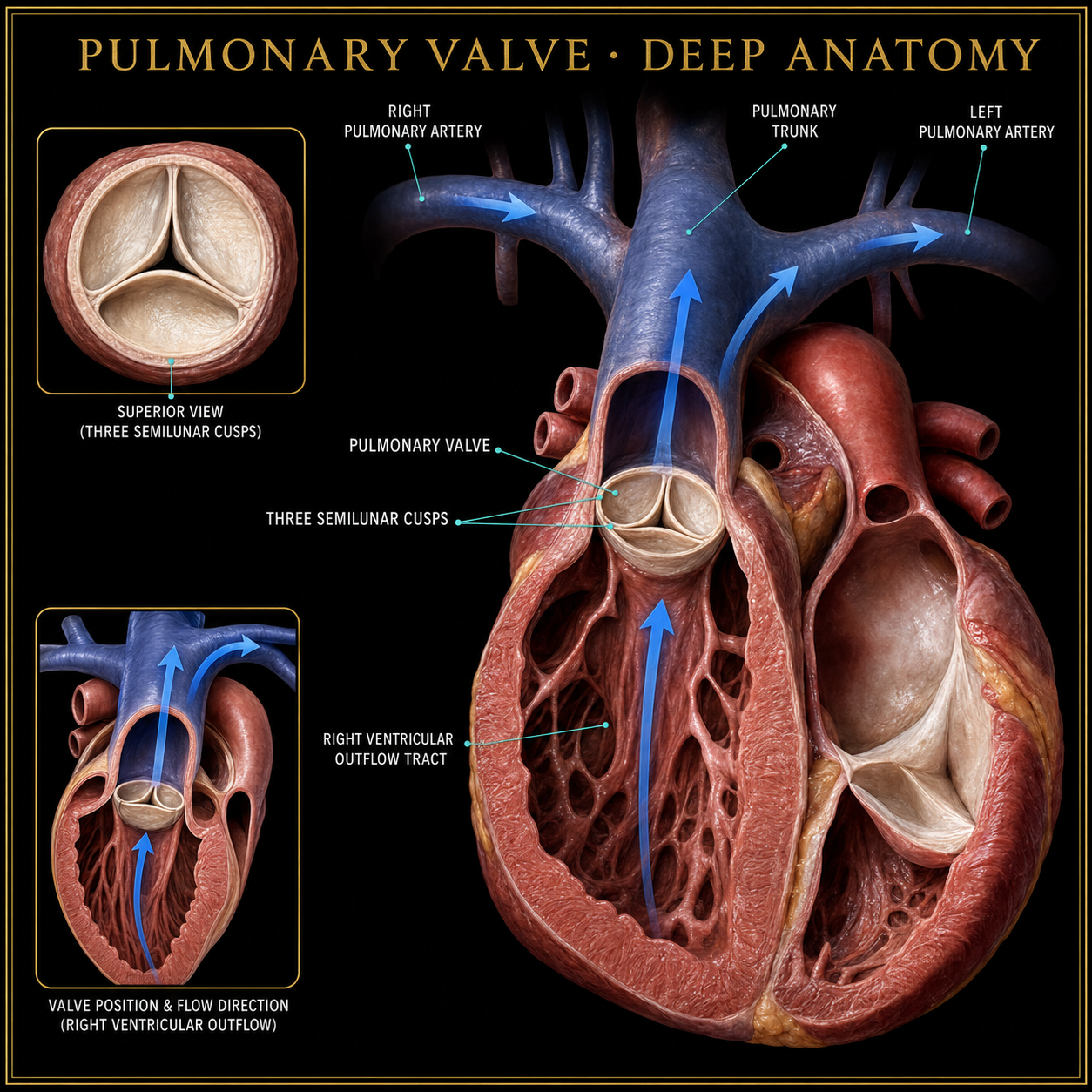 Open full image ↗
This semilunar valve opens between the right ventricle and pulmonary trunk, then closes to prevent blood from returning to the ventricle.
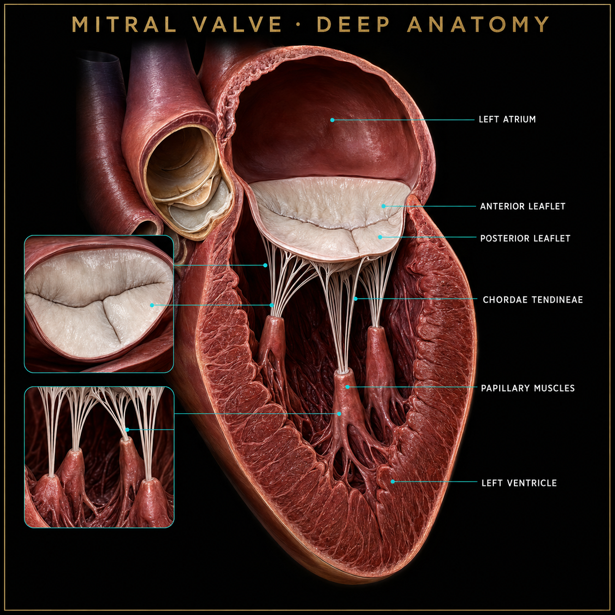 Open full image ↗
The left atrioventricular valve directs blood from the left atrium into the left ventricle.
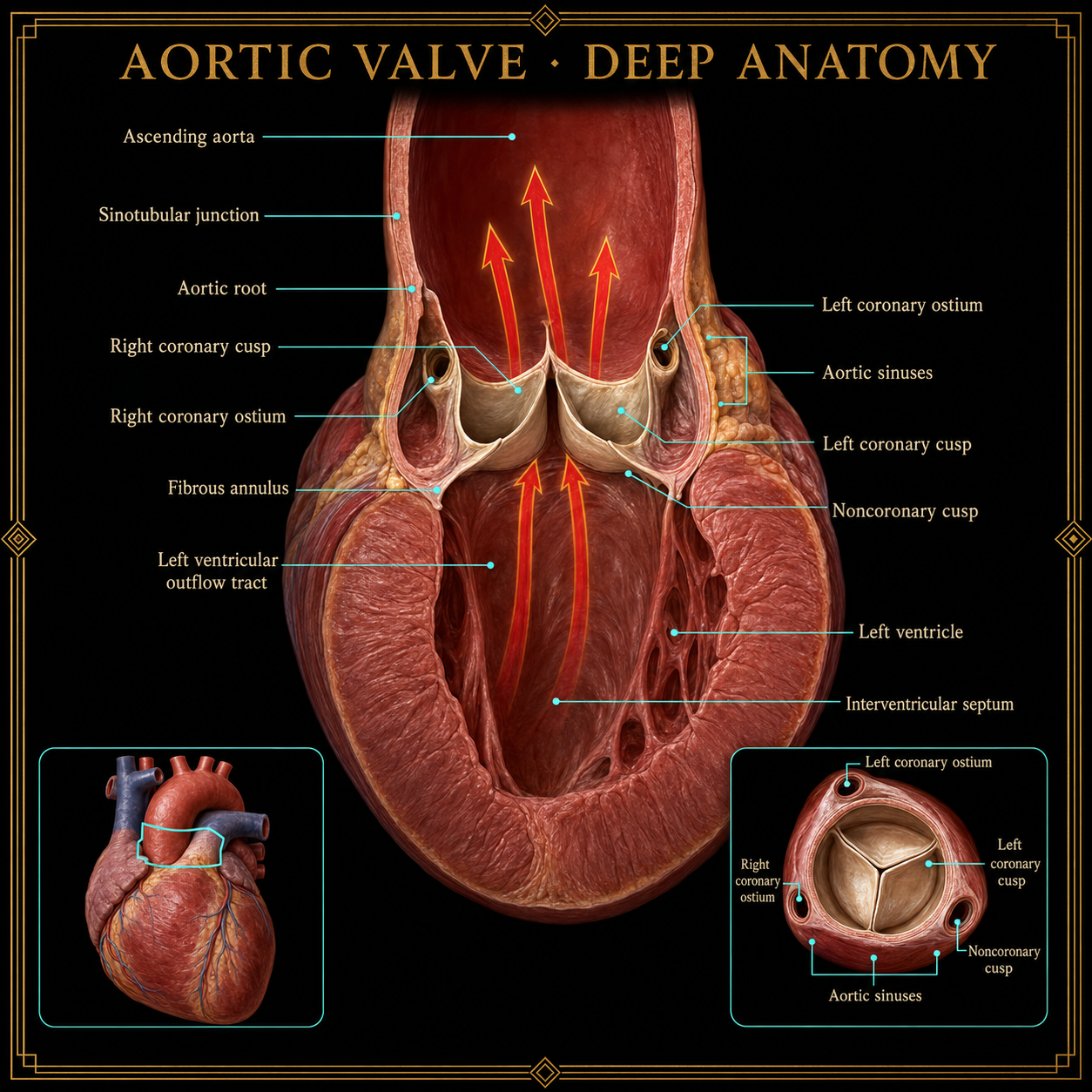 Open full image ↗
This semilunar valve opens between the left ventricle and aorta, allowing oxygenated blood to enter systemic circulation.
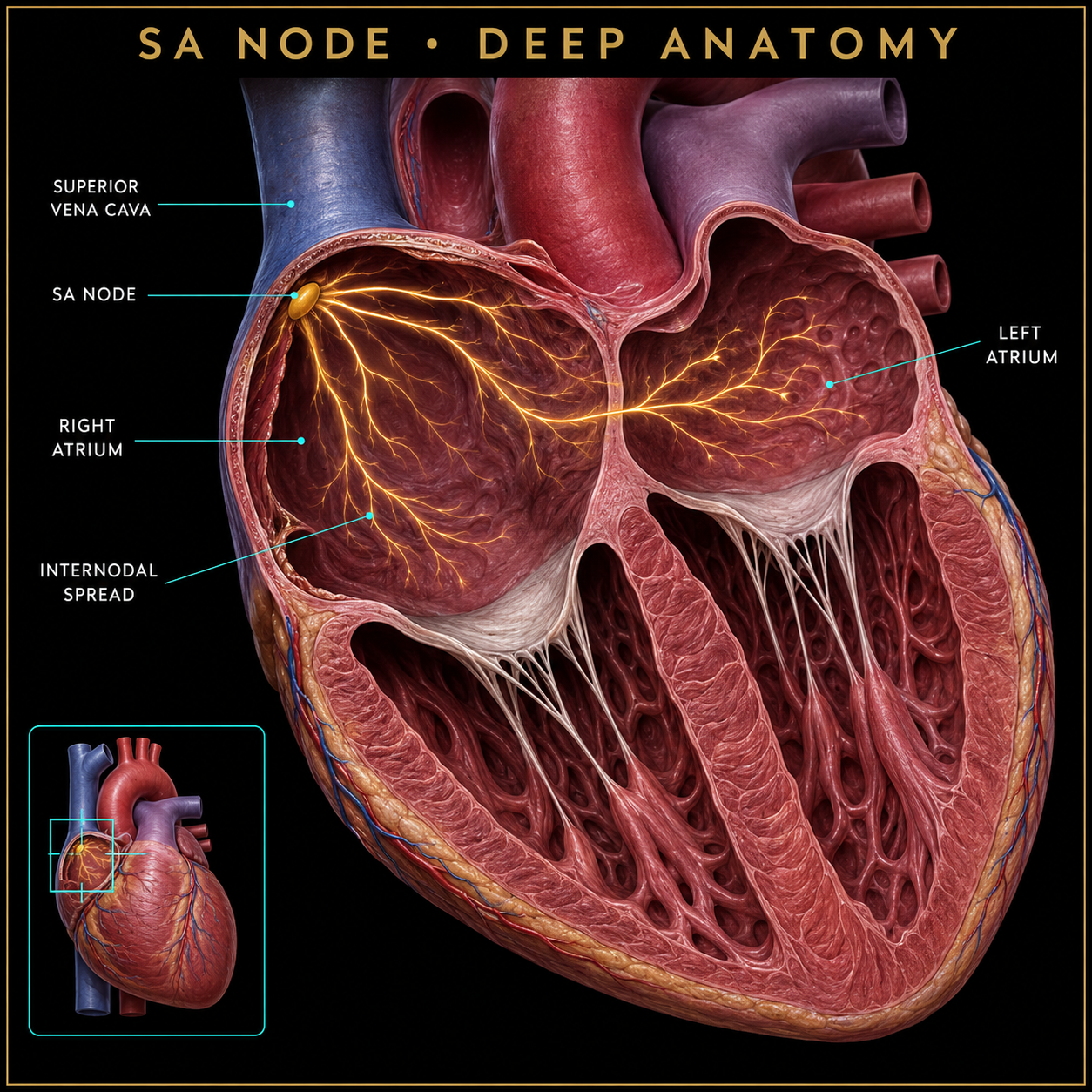 Open full image ↗
The sinoatrial node is the heart’s primary pacemaker. It initiates the electrical impulse that spreads across the atria.
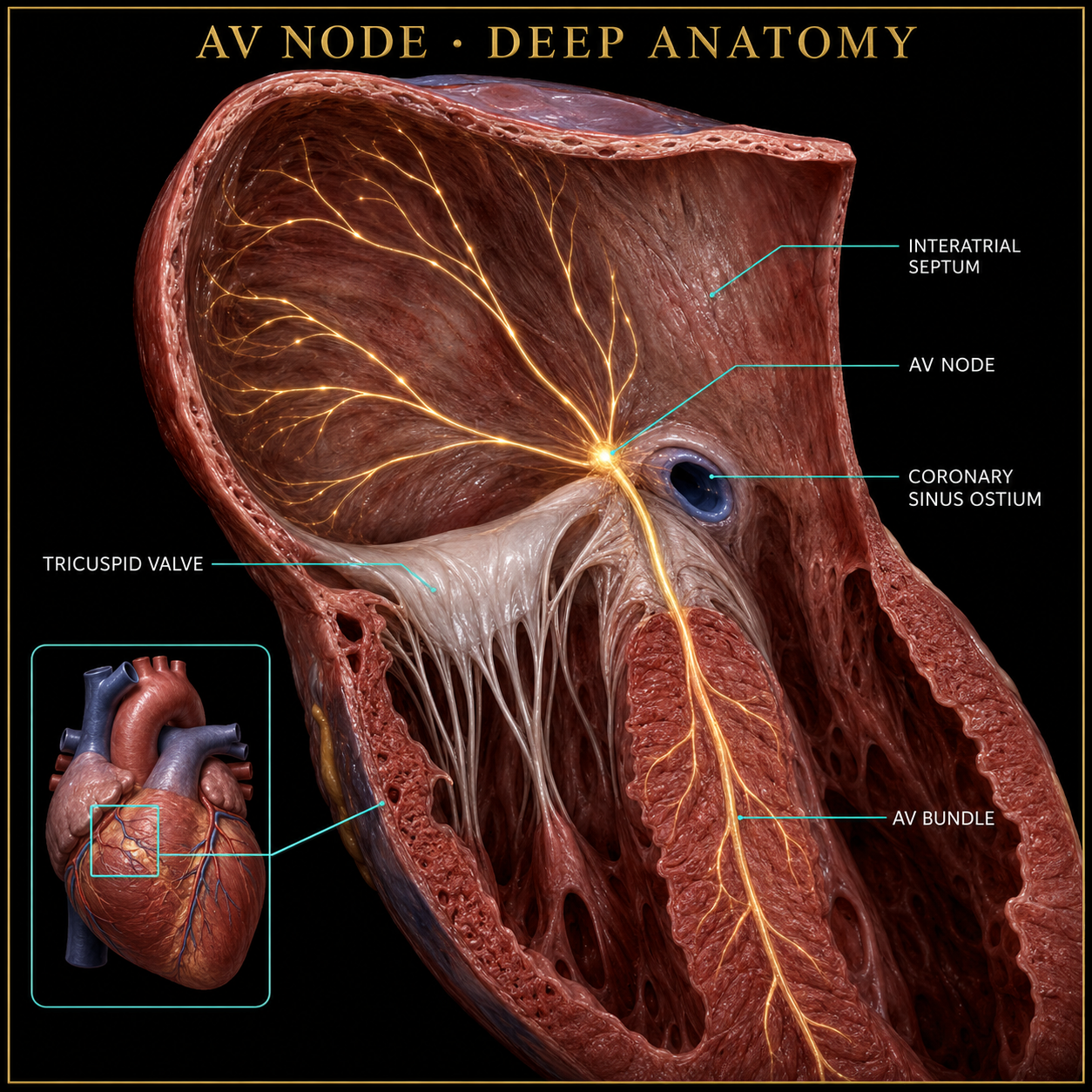 Open full image ↗
The atrioventricular node briefly delays the impulse so the ventricles have time to fill before they contract.
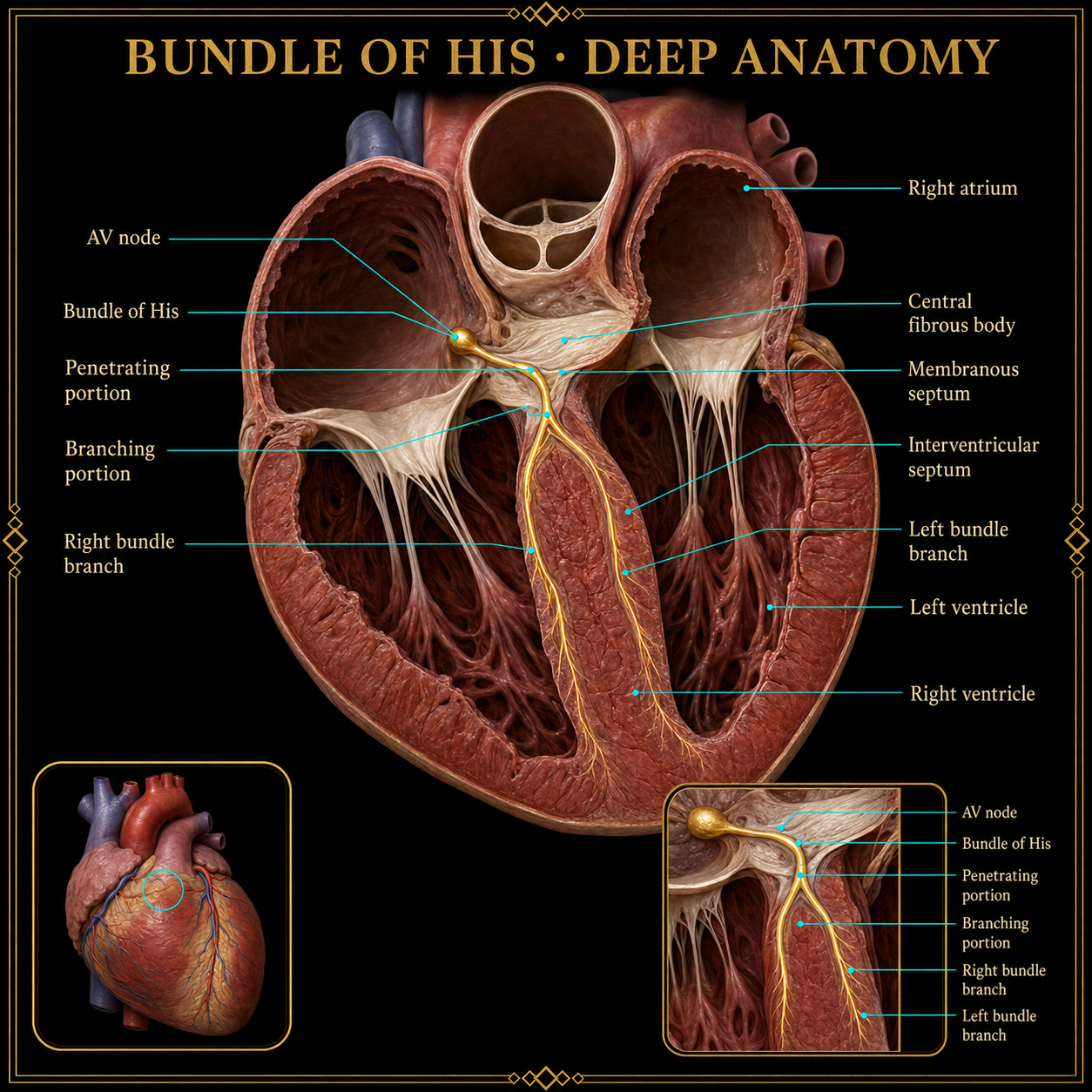 Open full image ↗
The atrioventricular bundle carries the impulse from the AV node through the heart’s fibrous skeleton into the upper interventricular septum.
 Open full image ↗
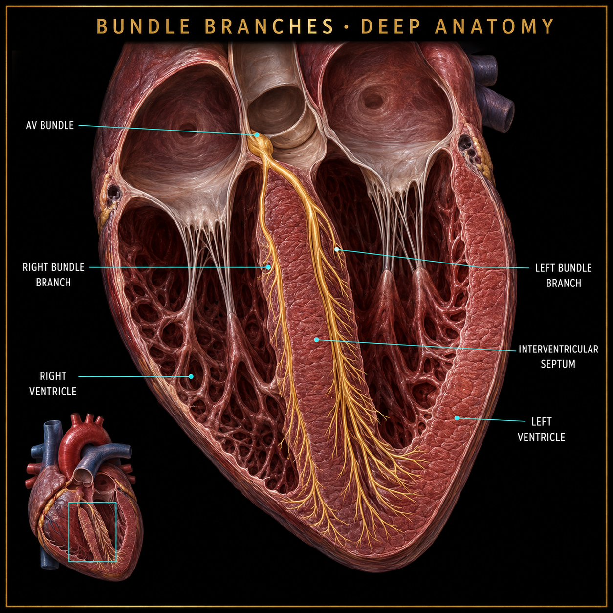
The right and left bundle branches split from the AV bundle and conduct the electrical impulse down the interventricular septum toward both ventricles.
 Open full image ↗
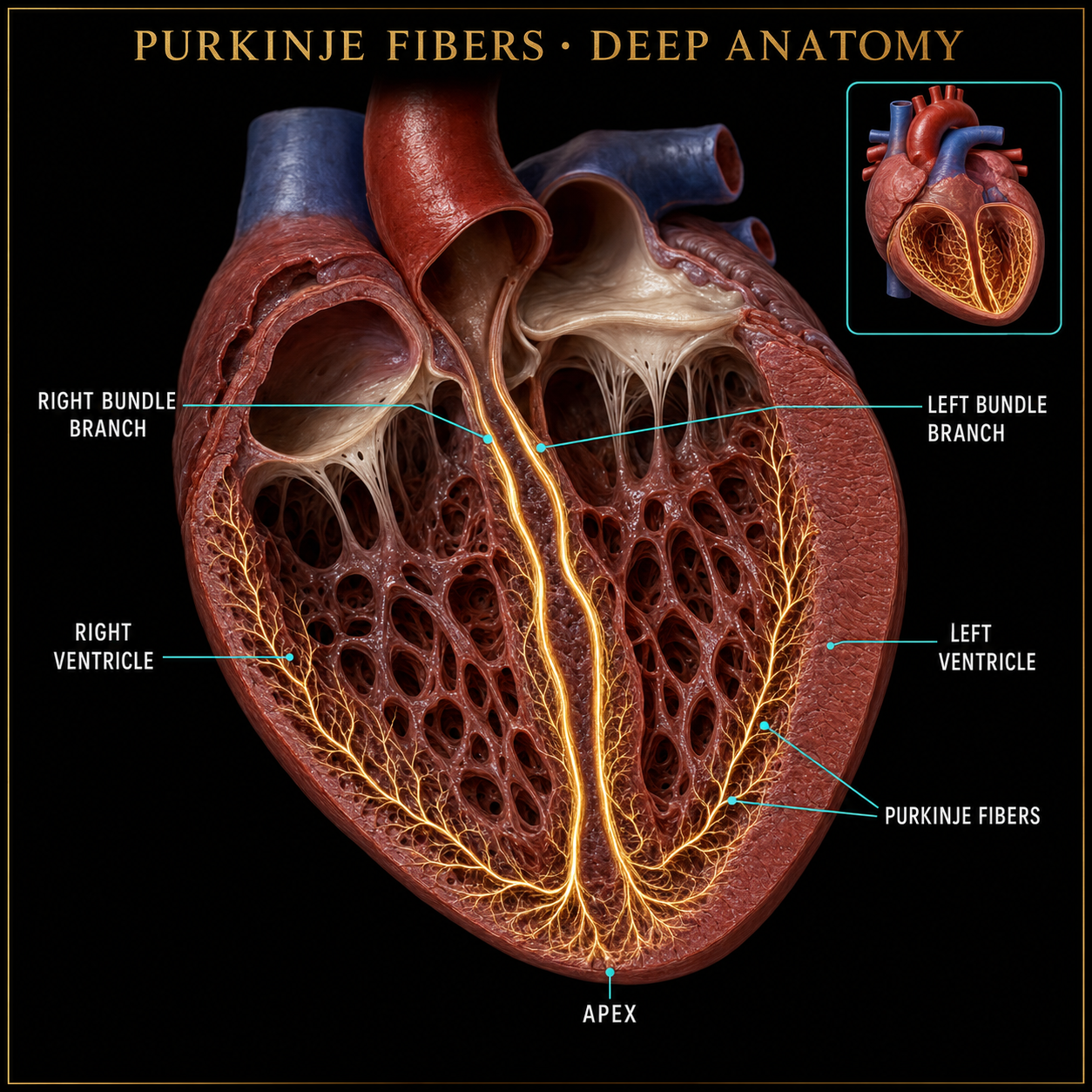
Purkinje fibers arise from the bundle branches and spread the electrical impulse through the myocardium of both ventricles.
 Open full image ↗
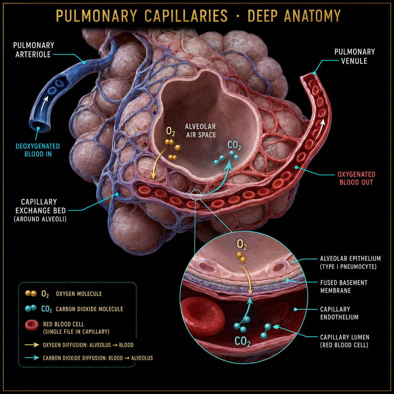
Capillaries surrounding the alveoli are the actual site where carbon dioxide leaves the blood and oxygen enters it.
 Open full image ↗
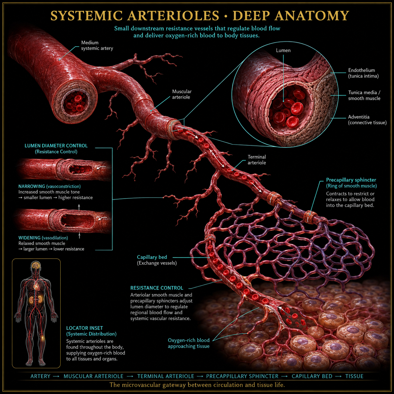
Small arterial branches downstream from arteries regulate resistance and deliver blood into systemic capillary beds.
 Open full image ↗
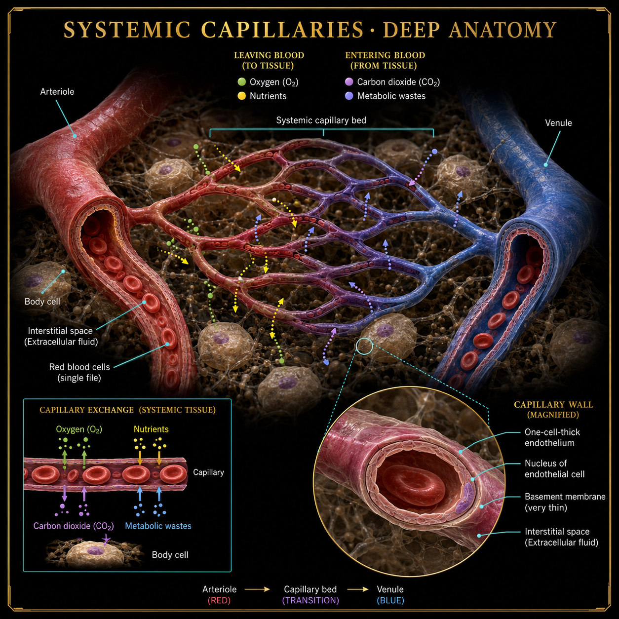
These microscopic exchange vessels deliver oxygen to tissues and receive carbon dioxide, nutrients and wastes.
 Open full image ↗
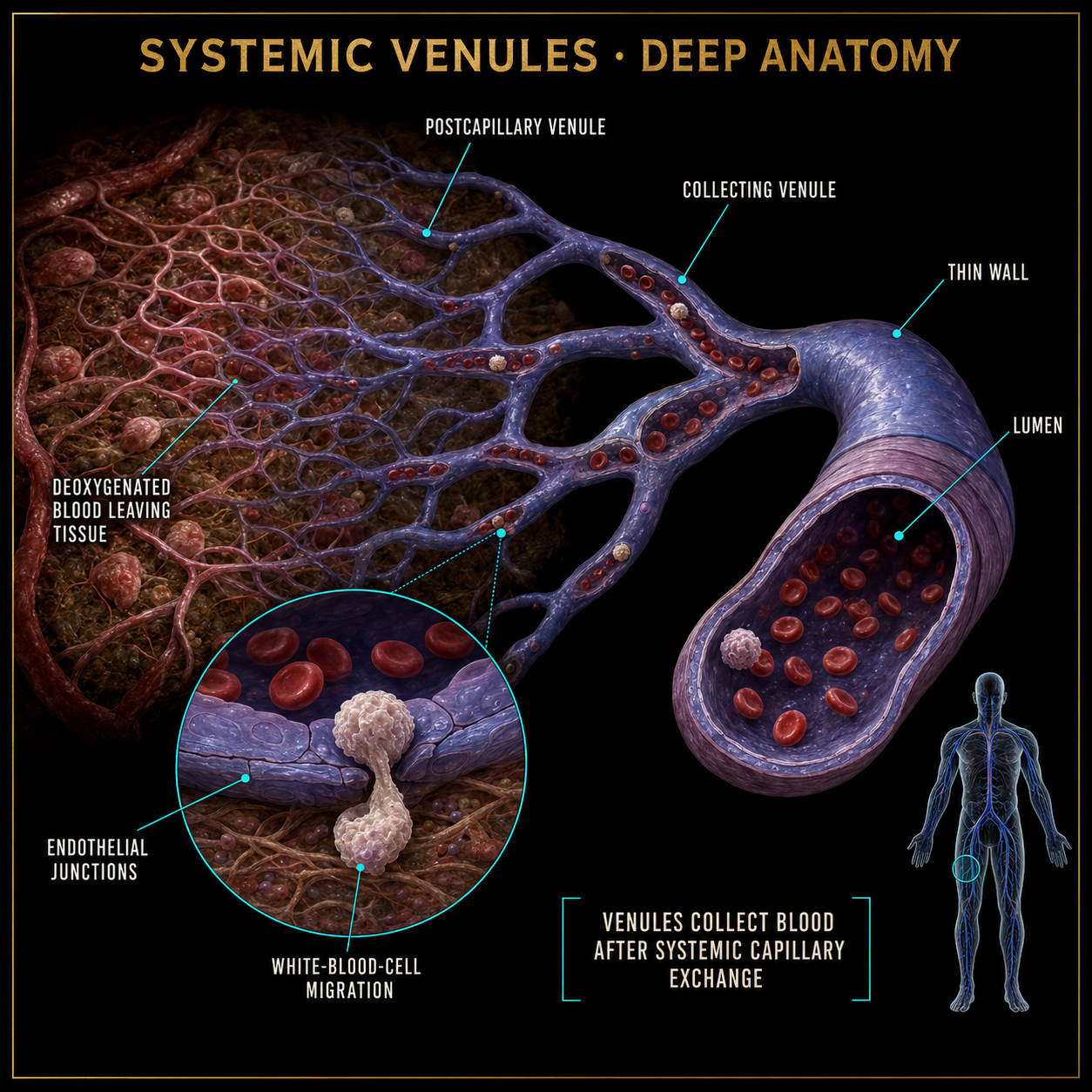
Venules collect oxygen-poor blood from systemic capillaries and merge into progressively larger veins.
 Open full image ↗
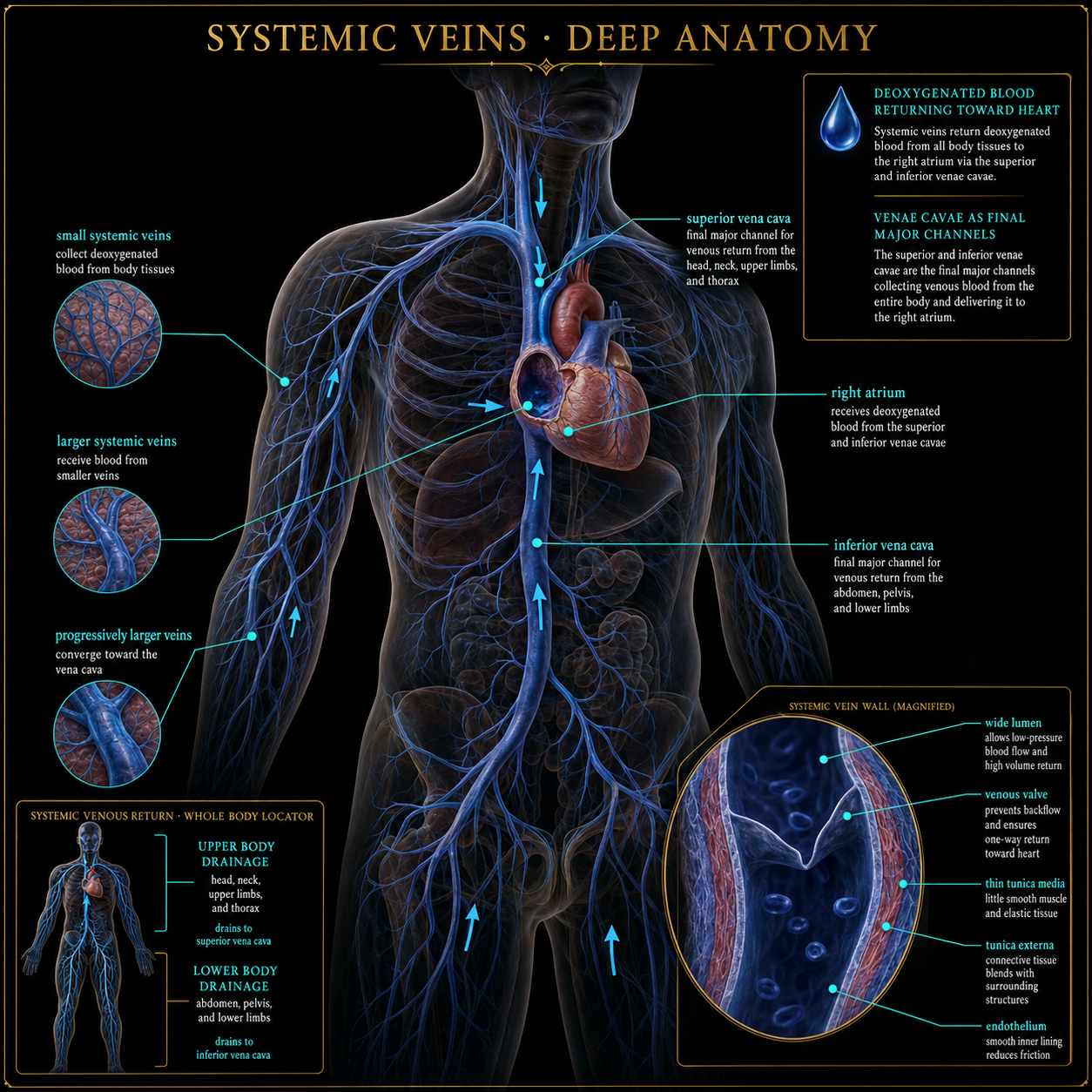
Systemic veins return blood toward the heart. Upper-body drainage reaches the superior vena cava; lower-body drainage reaches the inferior vena cava.
 Open full image ↗
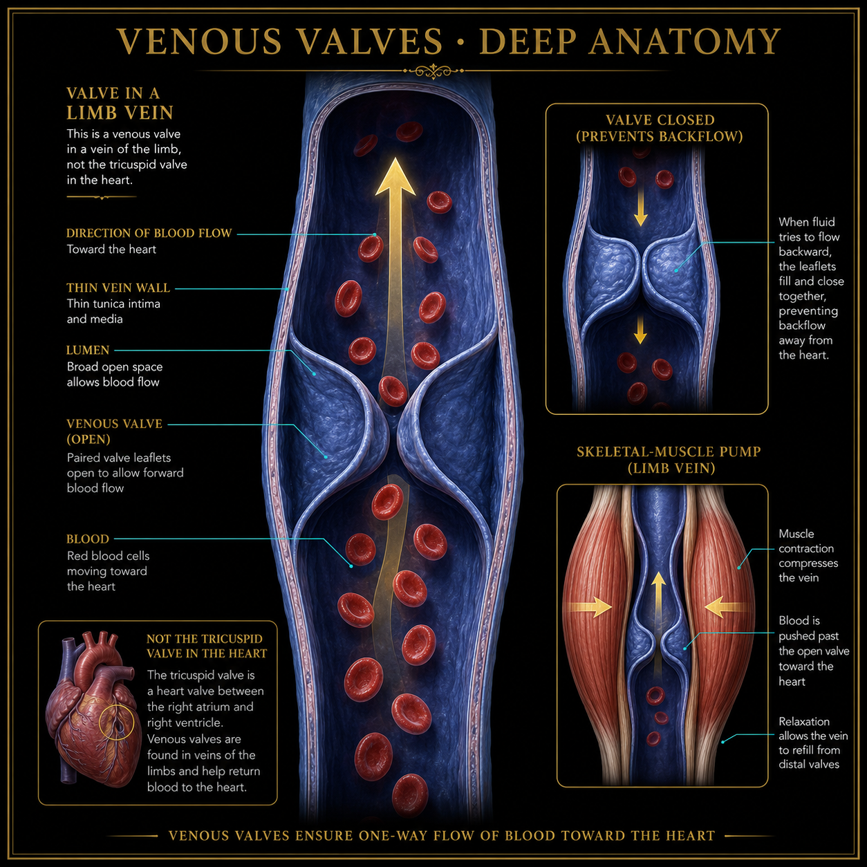
One-way valves within many veins—especially in the limbs—help prevent backflow. They are separate from the heart’s tricuspid valve.
 Open full image ↗
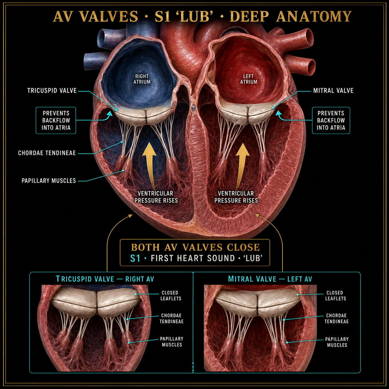
Closure of both atrioventricular valves—the mitral and tricuspid—is associated with the first heart sound, S1 or ‘lub.’
 Open full image ↗
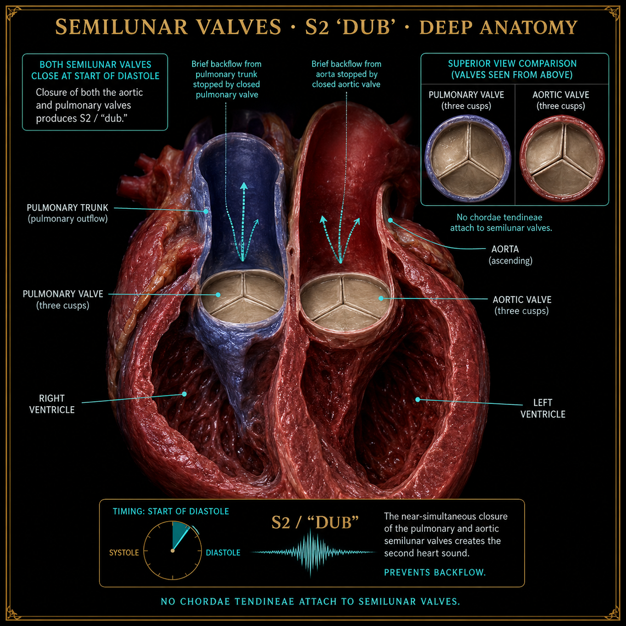
Closure of both semilunar valves—the aortic and pulmonary—is associated with the second heart sound, S2 or ‘dub.’
Educational artwork for the public beta. Anatomy plates will continue through medical-content review before formal classroom or clinical use.
Positive Vybez Only
See the Heart Lab turn into quick anatomy challenges, lyric breakdowns and behind-the-build clips.
Interactive 4D-motion public beta: time-synced lyrics, simulated depth, pulse and blood-flow overlays bring the realistic 2D anatomy scene to life. This is an educational prototype—not medical advice. Anatomy, timing and assessment content should be reviewed by qualified educators before formal classroom or clinical use.